Exploring SI Joint Anatomy and Its Function
The sacroiliac (SI) joint plays a pivotal role in our anatomy, yet it often goes unnoticed until it causes discomfort or pain. The SI joint is a complex structure that facilitates stability, mobility, and weight transfer between the spine and pelvis. Understanding SI joint anatomy can help us make sure it’s functioning properly and prevent potential injuries.
The Function of the SI Joint
The SI joint is a unique joint in the human body, comprising both synovial and fibrous components. Its intricate design allows it to fulfill multiple functions. When your SI joint functions incorrectly, it may cause pain and discomfort and lead to conditions such as sacroiliitis, sciatica, or piriformis syndrome. The following are some of the essential functions of the SI joint:
Weight Distribution
The SI joint is responsible for transferring the weight of the upper body to the lower extremities while standing, walking, or running. This weight-bearing function is essential for maintaining balance and stability. The SI joint anatomy is designed to withstand significant amounts of weight, making it one of the strongest joints in the body. The joint’s unique shape and connection to the pelvic bones allow it to distribute the weight evenly, reducing stress on other joints, such as the hips and knees.
Shock Absorption
Through its role in shock absorption, the SI joint reduces the impact of physical activities on the spine and other joints. During high-impact activities like running or jumping, forces are generated that may put significant stress on the body. With its robust ligaments and cartilage, the SI joint effectively absorbs these forces, reducing the pressure exerted on the spine and lower body.
This not only prevents potential injuries and wear and tear but also enhances comfort and endurance during these activities. Its capacity for shock absorption is a testament to the strength and resilience inherent in the SI joint anatomy.
Limiting Mobility
While the SI joint does allow for slight movements, its primary role is to provide stability to the pelvis and lower back. Excessive movement in this joint may lead to SI joint dysfunction and subsequent pain. The ligaments and muscles surrounding the SI joint help to limit its mobility, preventing potential injuries and maintaining proper alignment of the pelvis and spine. This is especially important during weight-bearing activities or when changing direction quickly.
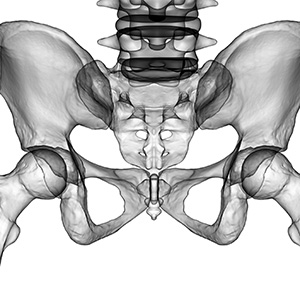
Understanding SI Joint Anatomy
The SI joint’s anatomy is as complex as its functions are diverse. It’s a hybrid joint, with one part being a synovial joint (the lower part) and the other a fibrous joint (the upper part). The articular surfaces are covered with a thin layer of hyaline cartilage on the sacral side and fibrocartilage on the iliac side. This unique structure allows the joint to manage significant load-bearing and stress.
SI Joint Structure
The structure of the SI joint is determined by the irregular surfaces of the sacrum and ilium bones that interlock with one another. Between these two bones, an extremely strong network of ligaments binds the joint together, providing structural support and limiting movement. These ligaments include the interosseous sacroiliac ligaments, the posterior sacroiliac ligaments, and the sacrotuberous and sacrospinous ligaments.
Muscles Supporting the SI Joint
Notably, several major muscles play a role in supporting the SI joint and ensuring its stability. The piriformis, gluteus Maximus, and hamstring muscles are key players in maintaining the joint’s stability. These muscles, in coordination with the strong ligaments, help control the minimal motion that occurs at the SI joint.
The Role of Cartilage Within the SI Joint Anatomy
The contribution of cartilage to the function and structure of the SI joint cannot be overlooked. The sacral side of the joint is covered with hyaline cartilage that measures approximately 1.18 mm in thickness, thicker than the iliac cartilage, which measures around 0.8 mm. The hyaline cartilage, renowned for its smooth and elastic properties, reduces friction in the joint and aids in the absorption of shock. On the other hand, the thinner iliac cartilage, despite being less robust, contributes to maintaining the overall stability of the joint. The cooperative work of these cartilages guarantees the smooth functioning of the SI joint, further highlighting the impressive complexity of SI joint anatomy.
What Is SI Joint Dysfunction?
SI joint dysfunction occurs when there is an abnormal increase or decrease in the joint’s mobility. This condition can cause pain in the lower back, hips, buttocks, and legs. If left untreated, it can significantly impact one’s quality of life. Various factors can contribute to SI joint dysfunction, including trauma, pregnancy, arthritis, and degenerative conditions. Identifying the root cause is essential to determine the most effective treatment approach. Symptoms of SI joint dysfunction may include:
- Pain and stiffness in the lower back, hips, buttocks, or legs
- Difficulty standing from a seated position
- Radiating pain down one or both legs
- Difficulty walking long distances or climbing stairs
Explore SI Joint Treatment With PainTEQ
PainTEQ’s LinQ SI Joint Stabilization System is a cutting-edge procedure designed to provide long-lasting SI joint pain relief by stabilizing the dysfunctional joint. Patients can contact PainTEQ to find a provider near them who specializes in diagnosing and treating SI joint dysfunction.
Physicians can partner with PainTEQ to begin offering this revolutionary procedure in their practice and discover how our technology can help your practice unlock success in SI joint treatment. Contact us today to learn more.