Types of SI Joint Provocative Tests for Diagnosing SI Joint Dysfunction
Sacroiliac (SI) joint dysfunction is a significant cause of lower back pain, yet it often remains underdiagnosed. Understanding the diagnostic process, particularly the SI joint provocative tests, is crucial for patients experiencing this type of pain. These tests are designed to replicate the pain and symptoms associated with SI joint dysfunction, aiding healthcare providers in pinpointing the source of discomfort.

Understanding SI Joint Dysfunction
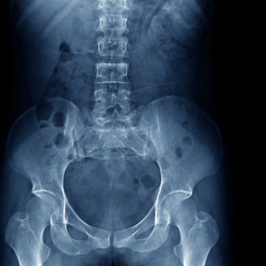
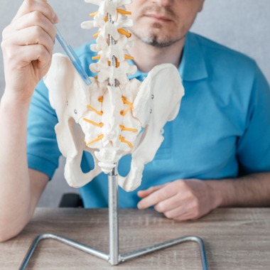
The SI joint connects the sacrum with the ilium (iliac bone) in the posterior pelvis, acting as a shock absorber and transmitting forces to the hips and lower extremities. Dysfunction in this joint can result from various causes, including traumatic events like falls or atraumatic factors such as pregnancy or infections.
Key SI Joint Provocative Tests
Healthcare providers use a combination of specific physical exam maneuvers, known as provocative tests, to diagnose SI joint dysfunction. Typically, a diagnosis is considered when at least three out of five tests are positive, with either the thigh thrust or compression test being one of them.
The following images and descriptions of SI joint provocative tests are sourced or informed by insights from a comprehensive review titled “Successful Diagnosis of Sacroiliac Joint Dysfunction,” published in the Journal of Pain Research. This article provides a detailed exploration of the diagnostic process for SI joint dysfunction, emphasizing the importance of these specific tests in identifying the condition.
1. FABER Test (Patrick’s Test)
The FABER test involves the patient lying supine, with the hip joint brought into a specific position and pressure applied to the knee and opposite hip. Pain in the SI joint area during this test suggests dysfunction.
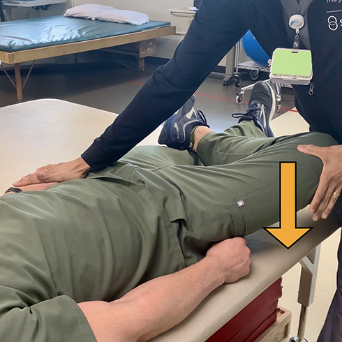
2. Thigh Thrust Test (Posterior Shear Test)
This test is performed with the patient lying supine, and an anterior-to-posterior shear force is applied to the SI joint through the femur. Pain at the SI joint indicates a positive test.
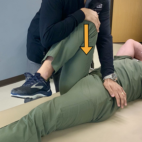
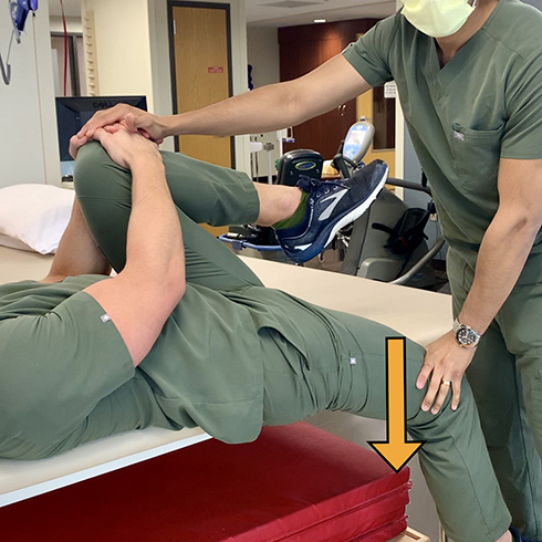
3. Gaenslen Test
In the Gaenslen test, the patient lies close to the table edge, with one leg hanging over and the other flexed to the chest. Pressure applied to both knees stresses the SI joints, and pain during this test is indicative of dysfunction.
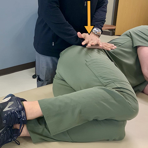
4. Compression Test
With the patient in a lateral position, downward pressure is applied to the iliac crest and ASIS. Pain in the SI joint on the affected side suggests a positive test.
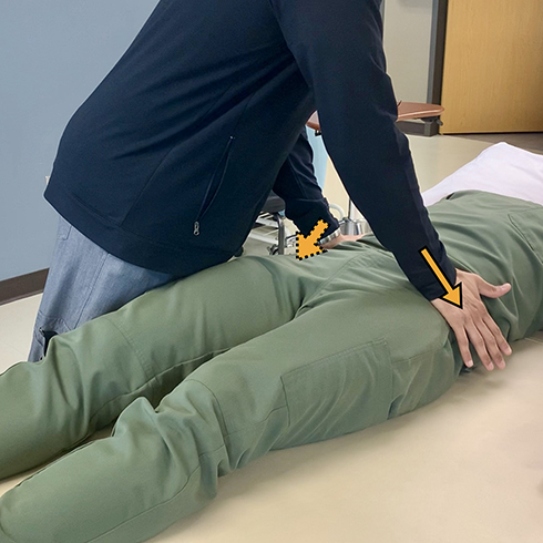
5. Distraction Test
This test involves applying outward pressure to the left and right ASIS with the patient lying supine. Pain in the SIJ area during this test signals dysfunction.
The Diagnostic Process
No single physical exam maneuver is diagnostic on its own. A combination of these tests, along with a detailed history and review of imaging, forms the basis of an accurate diagnosis. In the absence of pathognomonic tests, diagnostic SIJ blocks have evolved as the diagnostic standard.
Next Steps After Diagnosis
If you’ve been diagnosed with SI joint dysfunction, various treatment options are available. PainTEQ, known for its innovative LinQ SI Joint Stabilization System, can connect you with a partnering provider skilled in diagnosing SI joint dysfunction and determining if you’re a candidate for the LinQ procedure.
Understanding the diagnostic process, including SI joint provocative tests, is an essential step in addressing your pain effectively. If you suspect SI joint dysfunction, click here to connect with PainTEQ, who can help you find a physician near you.

